Images provide insight
Cone Beam Computed Tomography (CBCT) is a compact, faster and safer version of the regular CT. Through the use of a cone shaped X-Ray beam, the size of the scanner, radiation dosage and time needed for scanning are all dramatically reduced.
The time needed for a full scan is typically under one minute and the radiation dosage is up to a hundred times less than that of a regular CT scanner.
We use an advanced dental imaging system to produce high quality three-dimensional (3D) images that assist us in providing quality dental care. These scans provide high-definition 3D images of patients’ jaws and teeth. As a result, we get valuable information to help diagnose and solve your dental concerns. This is useful in the evaluation and placement of implants, bone pathology, as well as to plan for wisdom teeth extractions.
How Does It Work?
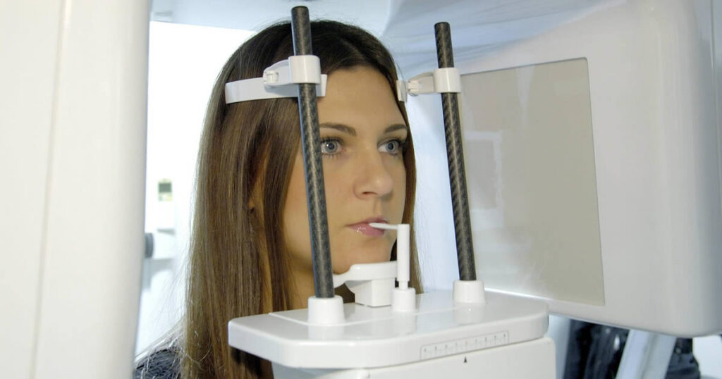
The 3D ConeBeam Imaging system is basically a digital x-ray scanner mounted on a rotating arm. (Like a digital camera, it uses digital technology to record images instead of old-fashioned film.) It’s called “ConeBeam” because the scanner projects x-rays in a carefully controlled, cone-shaped beam.
You simply sit in a chair while the scanner moves in one complete circle around your head, gathering all the scan data needed. There’s no special preparation necessary.
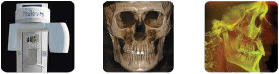
Your specialists can then call up whatever views they need on a computer monitor: 2D, 3D, “panoramic” views of your entire mouth, even sequences like pages in a book. They can view your images from any angle and in different magnifications, to see the relationships between bones, teeth, airways, nerves and tissues; to plan or evaluate your treatment.
And while 3D ConeBeam Imaging produces the same kind of high-quality images as a CT (CAT) scan, it does so with less radiation.
How Is It Different from Ordinary Dental X-rays?
Typical dental x-rays just focus on your teeth, and for each x-ray picture, you need one exposure. So it would take many exposures to even begin to compare to a single 3D ConeBeam scan. But 3D ConeBeam Imaging shows much more than simple “flat” x-rays. This new technology provides more complete visual information to study your case from every angle. Best of all, the original scan data can be duplicated anytime, to provide different specialists with images if needed later. And, there’s no film to get lost.
Why Do I Need It?
Whether you’re just beginning treatment or evaluating the results, 3D ConeBeam x-ray images give your doctors more of the high quality, detailed visual information they want for diagnosis and planning. More complete information is a vital key to improved patient care. And for you, it can mean more confidence and satisfaction in your treatment.
More Information
- No pre-scan preparation required
- Quick, simple, completely painless
- A virtually infinite number of views from just one 10-second scan
- Lower radiation dose than conventional CT scans
- Gives you the confidence that your doctor has advanced, precise information to plan and monitor your treatment